סריקה בתהליך...
הניתוח עשוי לארוך 30–90 שניות
הידעת?
הסרטון אינו מכיל תוכן רפואי
חושבים שחלה טעות?
אם לדעתכם הסרטון אכן עוסק בטענות רפואיות או בריאותיות, הוסיפו הסבר קצר ושלחו בקשה לבדיקה מחדש.
הבקשה התקבלה! נבדוק את הסרטון ונחזור אליכם.
אירעה שגיאה בשליחת הבקשה. נסו שוב.
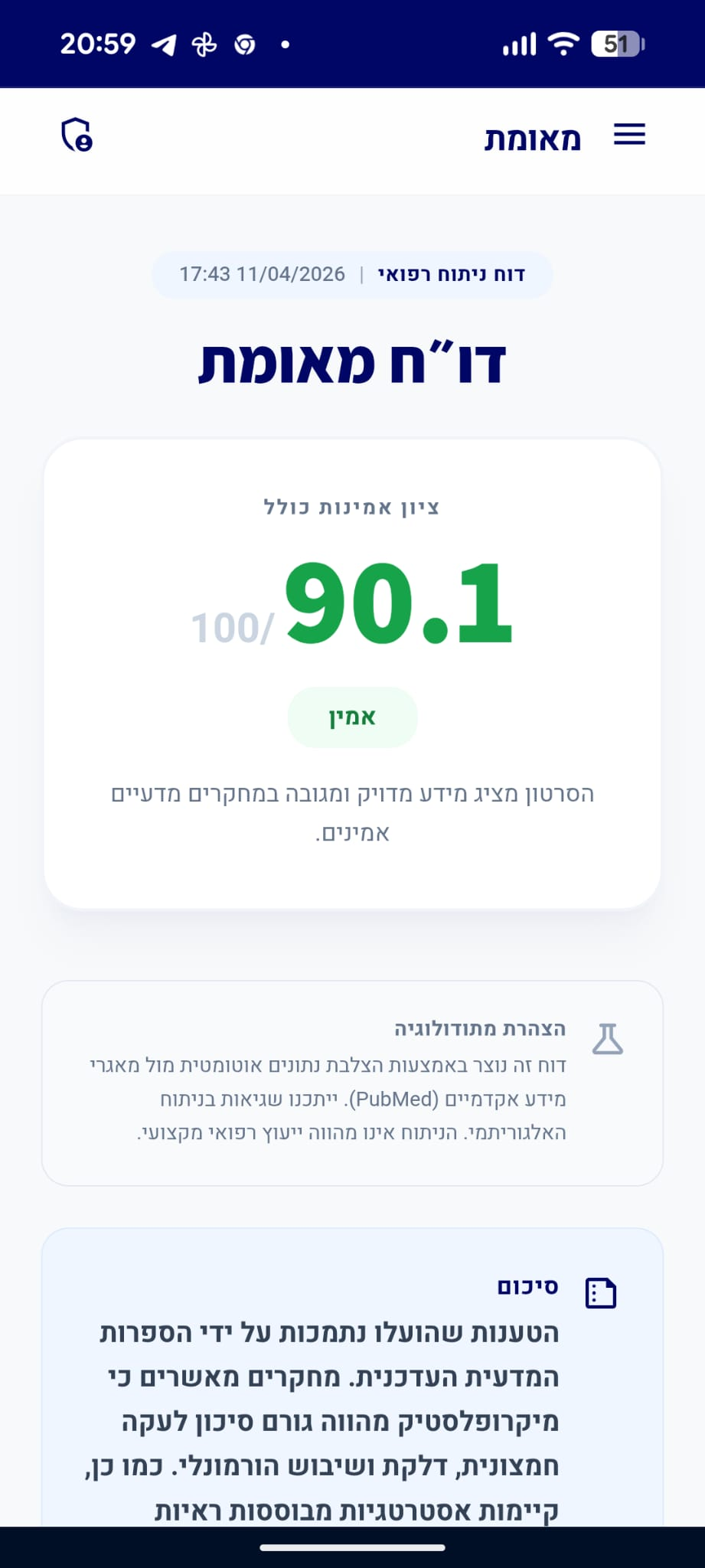
דו״ח מאומת
הסרטון מציג מידע מדויק ומגובה במחקרים מדעיים אמינים.
סיכום
המומחה הסביר את המנגנון הפיזיולוגי של נזקי השמש לעין, תוך הדגשה כי היעדר קולטני כאב ברשתית מונע מהאדם לחוש בנזק בזמן אמת. המידע שהציג המומחה תואם במדויק את הספרות המדעית העדכנית המאשרת כי חשיפה ישירה לשמש גורמת לרטינופתיה סולארית ולהופעת סקוטומות. לאור זאת, המלצתו של המומחה להימנע מהסתכלות ישירה על השמש היא נכונה ומבוססת מדעית.
דוח על סרטון תגובה
סרטון זה מציג קליפ של אדם המביע טענות רפואיות, ומומחה/מגיב שמתייחס אליהן. הציון מבוסס רק על טענות המומחה.
quiz טענות הקליפ ותגובת המומחה
"אנשים שמים מסך על העיניים מפני השמש"
"אל תסתכלו ישירות על השמש"
המומחה מספק המלצה רפואית למניעת נזק לעיניים.
מסקנת הבדיקה:
ההמלצה להימנע מהסתכלות ישירה על השמש היא קונצנזוס רפואי מוחלט המבוסס על הוכחות רבות לנזק בלתי הפיך או זמני לרשתית. (🟩)
chevron_right מקורות מדעיים: (3)
-
link
A Case of Bilateral Macular Phototoxicity and the Role of Multimodal Imaging.
Macular phototoxicity is a rare form of retinal injury caused by intense light exposure, most commonly from direct sun-gazing or viewing a solar eclipse without adequate eye protection. We report a case of bilateral macular phototoxicity in a 35-year-old female photographer with no significant past medical history, who developed acute central vision loss after approximately one hour of inadvertent sun exposure. She presented 48 hours after the incident with markedly reduced central vision (only light perception centrally) in both eyes, while peripheral vision remained intact. Dilated fundus examination revealed bilateral circumscribed focal foveal hypopigmentation in the absence of other retinal or optic nerve findings. Optical coherence tomography (OCT) scans confirmed foveal phototoxic damage, showing a normal overall contour but with juxtafoveal microcystic changes and hyperreflective lesions at the fovea in both eyes, consistent with acute macular phototoxicity. Conservative management with lubricating eye drops and oral nutritional supplements was implemented. Over the ensuing weeks, her visual acuity improved from an initial profound central scotoma to 20/40 in both eyes at 10 days, and 20/40 (left eye) and 20/30 (right eye) by one month post-exposure. Follow-up OCT demonstrated resolution of macular swelling and reconstitution of retinal layers. By five months, her visual acuity had fully recovered to baseline (20/20); however, a small persistent central scotoma remained in the left eye. This case illustrates the clinical course of macular phototoxicity and highlights the importance of patient education on ocular sun safety. Early recognition is important, as most cases show significant spontaneous recovery, but preventative measures are paramount since treatment options are limited.…
PMID: 41573477
-
link
The ophthalmic fallout in Utah after the Great American Solar Eclipse of 2017.
<h4>Background</h4>Solar retinopathy is a rare ophthalmic disorder resulting from sustained viewing of the sun without protective eyewear. Incidence of solar retinopathy typically increases following a solar eclipse due to attempted visualization of the sun without appropriate eye protection. This paper serves as a case series of all available reported cases of solar retinopathy present in Utah resulting from the August 21st, 2017 solar eclipse.<h4>Methods</h4>Twenty-seven patients had presented with concern for vision changes after the solar eclipse and six patients had exam findings consistent with solar retinopathy. Of these six cases, charts were available for three.<h4>Results</h4>The common finding in all cases was a central scotoma with a correlating change to the inner segment/outer segment junction on optical coherence tomography. Demographically, all three patients were young males.<h4>Conclusion</h4>This data provides insights on populations in Utah at risk for ophthalmic damage and can aid in targeting education programs in the future.…
PMID: 30275682
-
link
Solar retinopathy: A literature review.
Solar retinopathy (SR) refers to retinal injury that results from unprotected excessive exposure to light. It has been associated with direct sungazing, sunbathing, laser pointers, and welding arc exposure. Symptoms are typically bilateral and are characterized by asymmetric decreased vision, central or paracentral scotoma, photophobia, metamorphopsia, and headache. In most cases, recovery occurs spontaneously with no specific treatment within weeks to 6 months after exposure. However, few cases have been reported in the literature using steroids in acute SR because of their anti-inflammatory effects. The aim of this review is to present an update about this entity, describing the pathogenesis, risk factors, and diagnostic methods, with focus on management and outcomes of SR.…
PMID: 39132123
"כשאתה מסתכל ישירות על השמש, אתה מרכז קרינה ישירות לתוך אזור מסוים שנקרא מקולה"
המומחה מסביר את המנגנון הפיזיולוגי של הנזק הנגרם מהסתכלות ישירה על השמש.
מסקנת הבדיקה:
הספרות המדעית מאשרת כי הסתכלות ישירה על השמש גורמת לריכוז קרינה במקולה (מרכז הראייה ברשתית), מה שמוביל לנזק פוטוטוקסי המכונה רטינופתיה סולארית. (🟩)
chevron_right מקורות מדעיים: (3)
-
link
RETINAL BURNS IN PHOTIC RETINOPATHY: THREE CASE REPORTS.
Photic retinopathy (PR) is due to retinal phototoxicity, especially affecting the macula, resulting from exposure to sun, welding devices and lasers. It leads to oxidative damage to the retinal pigment epithelium (RPE) and the surrounding photoreceptors. Early recognition of this visual threatening condition, follow-up lesion evolution, and prevention of prolonged ocular exposure to lights is warranted. We herein report the three principal types of retinal burns due to solar retinopathy, laser pointer-induced maculopathy and arc welding maculopathy. La rétinopathie photique (RP) est secondaire à une phototoxicité rétinienne, affectant particulièrement la macula, résultant de l’exposition au soleil, aux appareils de soudure et aux lasers. Elle entraîne un dommage oxydatif de l’épithélium pigmentaire de la rétine (EPR) et des photorécepteurs. Il s’avère primordial de reconnaître précocement cette affection menaçant la vision, de suivre l’évolution des lésions et de prévenir une exposition oculaire prolongée aux lumières. Nous rapportons ici les trois principaux types de brûlures rétiniennes, dues à la rétinopathie solaire, à la maculopathie induite par un pointeur laser et à la maculopathie de soudure à l’arc.…
PMID: 39350887
-
link
A Case of Bilateral Macular Phototoxicity and the Role of Multimodal Imaging.
Macular phototoxicity is a rare form of retinal injury caused by intense light exposure, most commonly from direct sun-gazing or viewing a solar eclipse without adequate eye protection. We report a case of bilateral macular phototoxicity in a 35-year-old female photographer with no significant past medical history, who developed acute central vision loss after approximately one hour of inadvertent sun exposure. She presented 48 hours after the incident with markedly reduced central vision (only light perception centrally) in both eyes, while peripheral vision remained intact. Dilated fundus examination revealed bilateral circumscribed focal foveal hypopigmentation in the absence of other retinal or optic nerve findings. Optical coherence tomography (OCT) scans confirmed foveal phototoxic damage, showing a normal overall contour but with juxtafoveal microcystic changes and hyperreflective lesions at the fovea in both eyes, consistent with acute macular phototoxicity. Conservative management with lubricating eye drops and oral nutritional supplements was implemented. Over the ensuing weeks, her visual acuity improved from an initial profound central scotoma to 20/40 in both eyes at 10 days, and 20/40 (left eye) and 20/30 (right eye) by one month post-exposure. Follow-up OCT demonstrated resolution of macular swelling and reconstitution of retinal layers. By five months, her visual acuity had fully recovered to baseline (20/20); however, a small persistent central scotoma remained in the left eye. This case illustrates the clinical course of macular phototoxicity and highlights the importance of patient education on ocular sun safety. Early recognition is important, as most cases show significant spontaneous recovery, but preventative measures are paramount since treatment options are limited.…
PMID: 41573477
-
link
Solar retinopathy: a review of the literature and case report.
Accounts of solar retinopathy have existed for centuries, but only recently have researchers begun to investigate the mechanisms responsible for producing solar retinal injury. The vast majority of solar retinal injuries occur as a result of viewing a solar eclipse without adequate protection. The extent of structural retinal damage and associated visual impairment is dependent upon the intensity and duration of solar exposure. The exact mechanisms which operate to produce solar retinal compromise are not completely known, but is believed to involve a thermally enhanced photochemical process. Despite the lack of a standardized treatment protocol, most cases of solar retinopathy will improve significantly over time without treatment. Prevention remains the mainstay of therapy. Solar retinopathy must be differentiated from other subtle macular diseases in the absence of a confirmed history of solar exposure.…
PMID: 3998365
"ברשתית אין קולטני כאב, ולכן אתה אולי גם חושב שזה בסדר, או שלפחות אין נזק"
המומחה מסביר מדוע אנשים עלולים להמשיך להסתכל על השמש למרות הנזק המצטבר.
מסקנת הבדיקה:
הרשתית אכן חסרה קולטני כאב (נוציספטורים), מה שמסביר מדוע נזק פוטוכימי מצטבר יכול להתרחש ללא תחושת כאב מיידית שתתריע בפני המטופל על הסכנה. (🟩)
"אחרי כמה ימים או שעות אפילו, יתום יהיה לך איזה קטם בעין, שחור כזה"
המומחה מתאר את התוצאה הקלינית של הסתכלות ישירה על השמש.
מסקנת הבדיקה:
מחקרים קליניים מאשרים כי הסתכלות ישירה על השמש מובילה להופעת סקוטומה (נקודה עיוורת או כתם שחור בשדה הראייה) כתוצאה מנזק לרשתית, לעיתים תוך שעות או ימים מהחשיפה. (🟩)
chevron_right מקורות מדעיים: (3)
-
link
[Microperimetry and reading saccades in retinopathia solaris. Follow-up with the scanning laser ophthalmoscope].
Patients with solar retinopathy often complain of minute central scotomas and are handicapped when reading. The purpose of this study was to verify scotomas that are too small to be detected by standard perimeters and to analyze patients' reading patterns. Nineteen patients (12 female, 7 male, aged 5-46 years) with acute solar retinopathy after watching a solar eclipse on 12 October 1996 underwent scanning laser ophthalmoscope (SLO) microperimetry within 10 days after exposure using stimulus size Goldmann I (0.11 degree) with the 20 degrees field. Size and depth of scotomas were measured. Eye movements during reading were recorded on videotape. Follow-up was at 1 and 6 months. Thirty-one eyes (7 patients unilateral, 12 bilateral) showed scotomas. Four eyes showed anatomic changes in the retinal pigment epithelium but no functional loss. VA was 0.16 to 0.5 in 5 eyes, 26 had VA of 0.8-1.2. Scotomas could be detected in all eyes with subjective impairment. Scotoma size varied from 0.3 to 1.7 degrees (1 patient 6 degrees). Forty-four percent were deep scotomas (0 dB). All defects improved at 1 and at 6 months; 25% were no longer detectable. Reading speed was reduced in 75% of eyes (42% at 6 months): 200-560 signs/min, median: 510, normal > or = 660 (at 6 months: 350-920 signs/min, median 670). This was especially due to increased number of regressions (in 81% of eyes, 21% at 6 months). The frequency and width of saccades were no different from normal subjects. Minute scotomas (diameter = 0.3 degree) can be detected with the SLO. All patients showed objective improvement of their field defect up to 6 months, even when this was not noted by the patient or thought to be due to habituation. Small scotomas can dramatically reduce reading performance.…
PMID: 10414122
-
link
A Case of Bilateral Macular Phototoxicity and the Role of Multimodal Imaging.
Macular phototoxicity is a rare form of retinal injury caused by intense light exposure, most commonly from direct sun-gazing or viewing a solar eclipse without adequate eye protection. We report a case of bilateral macular phototoxicity in a 35-year-old female photographer with no significant past medical history, who developed acute central vision loss after approximately one hour of inadvertent sun exposure. She presented 48 hours after the incident with markedly reduced central vision (only light perception centrally) in both eyes, while peripheral vision remained intact. Dilated fundus examination revealed bilateral circumscribed focal foveal hypopigmentation in the absence of other retinal or optic nerve findings. Optical coherence tomography (OCT) scans confirmed foveal phototoxic damage, showing a normal overall contour but with juxtafoveal microcystic changes and hyperreflective lesions at the fovea in both eyes, consistent with acute macular phototoxicity. Conservative management with lubricating eye drops and oral nutritional supplements was implemented. Over the ensuing weeks, her visual acuity improved from an initial profound central scotoma to 20/40 in both eyes at 10 days, and 20/40 (left eye) and 20/30 (right eye) by one month post-exposure. Follow-up OCT demonstrated resolution of macular swelling and reconstitution of retinal layers. By five months, her visual acuity had fully recovered to baseline (20/20); however, a small persistent central scotoma remained in the left eye. This case illustrates the clinical course of macular phototoxicity and highlights the importance of patient education on ocular sun safety. Early recognition is important, as most cases show significant spontaneous recovery, but preventative measures are paramount since treatment options are limited.…
PMID: 41573477
-
link
Solar retinopathy: A literature review.
Solar retinopathy (SR) refers to retinal injury that results from unprotected excessive exposure to light. It has been associated with direct sungazing, sunbathing, laser pointers, and welding arc exposure. Symptoms are typically bilateral and are characterized by asymmetric decreased vision, central or paracentral scotoma, photophobia, metamorphopsia, and headache. In most cases, recovery occurs spontaneously with no specific treatment within weeks to 6 months after exposure. However, few cases have been reported in the literature using steroids in acute SR because of their anti-inflammatory effects. The aim of this review is to present an update about this entity, describing the pathogenesis, risk factors, and diagnostic methods, with focus on management and outcomes of SR.…
PMID: 39132123

Elay Cohen | לימודי רפואה בחו״ל
דירוג זה מבוסס על 9 דוחות אימות קודמים.

האם הדוח הזה היה מועיל לך?
מה היה פחות טוב? (רשות)
תודה על הפידבק!
עירעור על דוח זה
ספקו ראיות חדשות או הצביעו על אי דיוקים
נעדכן אותך על תוצאות הבדיקה
הוסיפו קישורים למחקרים או מקורות רפואיים מוכרים
העירעור נשלח בהצלחה!
המנוע המדעי שלנו יבדוק את הראיות שהגשתם. נעדכן אתכם באימייל עם התוצאות.
ניתוח מבוסס בינה מלאכותית
דוח זה נוצר באופן אוטומטי על ידי מערכת בינה מלאכותית ועשוי להכיל שגיאות, אי-דיוקים או מידע חלקי. הניתוח אינו מהווה ייעוץ רפואי, אבחנה או המלצה לטיפול, והוא אינו תחליף לדעתו של איש מקצוע רפואי מוסמך. יש להתייעץ עם רופא או מומחה מוסמך לפני קבלת כל החלטה רפואית. המידע מוצג לצרכי מידע כללי בלבד.
מידע זה מופק על ידי בינה מלאכותית ואינו מהווה תחליף לייעוץ רפואי מקצועי.